Valvular heart disease is any disease that involves one or more than one type of valves in the heart (aortic and mitral valve in the left ventricle, and the pulmonary and tricuspid valve in the right heart). Valvular heart disease is usually a disease congenital (inborn) or acquired (due to several reasons that occur after birth). Heart valve disease management may include medical treatment, but when it gets worse then it should be done surgery, such as valve repair or replacement (repair or replacement).
Some causes of heart valve abnormalities.
Dysplasia.
Valve dysplasia is a disorder of development that is at the heart valves (commonly exposed to one or more of the heart valves). The disorder often occurs is tetralogy of Fallot, where the abnormality is composed of four kinds of abnormalities with one of them is narrowing / stenosis of the pulmonary valve. Ebstein's anomaly is an abnormality of the tricuspid valve.Rheumatic Heart Disease.
Valvular heart disease caused by fever rhema called "rheumatic heart disease". In the developing countries (including Indonesia), the incidence of rheumatic heart disease is still very high, and diminishing in developed countries. It is closely linked with the number of infectious diseases and better treatment of infections in developed countries.Inflammatory.
Inflammatory / inflammation of the heart valves whatever the cause is called endocarditis; most often caused by a bacterial infection, commonly also from cancer (marantic endocarditis), or some autoimmune circumstances (Libman-Sacks endocarditis) and hypereosinophilic syndrome (Loeffler endocarditis). Some of the drugs associated with heart valve disease emergence is ergotamine such as pergolide and cabergoline.Mitral heart valve abnormalities.
Mitral valve (also known as the bicuspid valve / left atrioventricular valve) is the valve that is in the heart consists of two cusps. The mitral valve is a heart valve that separates anatara left atrium and left ventricle). The mitral valve and tricuspid valve is due to atrioventricular valve is located between the foyer and the heart chambers, and both control the blood flow rate.When diastole, normal functioning mitral valve opens as a result of increasing pressure from the left atrium fills with blood (preload). When the atrial pressure increases above the left ventricle, the mitral valve opens allowing blood to flow passively to the left ventricle. Diastole ended when 20% contraction of the atria pump blood from the rest of the left atrium to the left ventricle, which is referred to as end diastolic volume (EDV), and mitral valve closes at the end of atrial contraction to prevent blood flow back to the heart
Anatomy.
Mitral valve (bicuspid valve) It is in the heart (between the atrium and the left ventricle)The average size of mitral valve 4-6 cm². The mitral valve has two valve / flyers (leaflets anteromedial and posterolateral leaflet). Valve valve is limited by a ring called the annulus of the mitral valve. Valve covers 2/3 of the anterior mitral valve area, and the rest by the posterior valve. Valve is guarded by a tendon attached to the posterior part of the valve, prevent valve prolapse. These tendons called chordae tendineae.
Chordae tendineae to the papillary muscles attached to its end (papillary muscles) and the valve. Papillary muscle itself is a protrusion of the left ventricular wall. When the left ventricle contracts, intraventricular pressure forced the mitral valve to close. Tendon keep the leaflet remain parallel to each other and do not leak into the atrium.
Normal physiology.
When the left ventricular diastole, after the reduced pressure in the left ventricle due to muscle relaxation ventricle, the mitral valve opens and blood flow from the left atrium into the ventricle kiri.sebanyak 70-80% of the blood flowing through the early phase of filling of the left ventricle (move because of the pressure difference).Contraction of the left atrium (left atrial systole) simultaneously with left ventricular diastole, causing the rest of the blood remaining in the left atrium immediately flows into the left ventricle. This is also referred to as atrial kick.
Annular / ring of the valve change shape and size when the cardiac cycle lasts. The shape shrink when atrial systole because of left atrial contraction. Disturbances in the annulus, valve and mitral valve support structure can make the mitral valve is leaking (regurgitation / insufficiency) or narrowed (stenosis).
General Operating Procedures.
The surgical procedure on heart disease is largely synonymous but will have different techniques at the core of its operations, depending on the abnormalities that occur.
Most operations will use a heart-lung machine in the procedure, but there are also techniques that do not use the heart-lung machine for example at Off Pump CABG, where the heart will continue beating during surgery.
Measures heart surgery commonly performed are as follows :
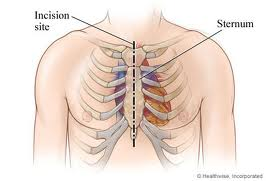
- Physician Anesthesia will perform general anesthesia.- Surgeon will start an incision in the midline of the chest, from the skin to open the breastbone. At BPAK surgery, previous surgeon will take the blood vessels that will be installed in the heart, the saphenous vein in the leg or the radial artery in the arm.
- Then the chest cavity will be widened by using a retractor.
- If the heart-lung machine is used, the heart will be connected to the heart-lung machine and cardioplegic substances included. Cardioplegic substances while making the heart stops beating and the heart-lung machine takes over the function of the vital organ.
- Operations carried out in accordance with the diagnosis of disease.
- Heart made pulsing back so that the heart-lung machine can be weaned.
- When the work of the heart and lungs are already stabilized, closed chest wall and sternum connected using a special wire.
Thank you for reading this article. Written and posted by Bambang Sunarno. sunarnobambang86@gmail.com
author:
https://plus.google.com/105319704331231770941.
name: Bambang Sunarno.
http://primadonablog.blogspot.com/2015/09/you-know-about-heart-valve-disease.html
DatePublished: 14 September 2015 12:32
Tag : Heart, Valve Disease, Heart Valve Disease,
Code : 7MHPNPADAEFW


No comments:
Post a Comment